- Motion Realizing
- info@neuromove.it

DIAGNOSTIC TECHNOLOGY AS A PREVENTIVE MEASURE AND ACTION PLAN

Palabras claves: #Medidaspreventivas , #diagnosticoporimágenes, #observacióncientífica, #movimiento, #trabajointerdisciplinario, #tecnología y #Neurociencias
Introduction:
Con la escritura de este articulos queremos mencionar las bases fisicas de la tegnologia que permite realizar diagnostico de lesiones, disfunciones y traumas.
Además establecer simples protocolos de accción preventiva, en pos de direccionar a nuestros alumnos/ pacientes / jugadores y amigos, luego haber sufrido una lesión, o simplemente por el sentimiento de dolor .
Historical review X-rays
Wilhem Conrad Röntgen, German physicist discovered X-rays in 1895, while developing an experiment with an empty tube and electrodes to generate high-voltage currents. He called it Crookes tube. This tube, being close to photographic plates, generated in them some blurred images. After covering the tube with black cardboard to eliminate visible light, he observed a faint yellow-green glow from a screen with a layer of platinum-barium cyanide, which disappeared when the tube was turned off. He determined that the rays created a very penetrating, but invisible, radiation that went through great thicknesses of paper and even thin metals. He used photographic plates to demonstrate that the objects were more or less transparent to X-rays depending on their thickness and he made the first human X-ray, using his wife's hand. I call them
Wilhem Conrad Röntgen, German physicist discovered X-rays in 1895, while developing an experiment with an empty tube and electrodes to generate high-voltage currents. He called it Crookes tube. This tube, being close to photographic plates, generated in them some blurred images. After covering the tube with black cardboard to eliminate visible light, he observed a faint yellow-green glow from a screen with a layer of platinum-barium cyanide, which disappeared when the tube was turned off. He determined that the rays created a very penetrating, but invisible, radiation that went through great thicknesses of paper and even thin metals. He used photographic plates to demonstrate that the objects were more or less transparent to X-rays depending on their thickness and he made the first human X-ray, using his wife's hand. I call them
Wilhem Conrad Röntgen, German physicist discovered X-rays in 1895, while developing an experiment with an empty tube and electrodes to generate high-voltage currents. He called it Crookes tube. This tube, being close to photographic plates, generated in them some blurred images. After covering the tube with black cardboard to eliminate visible light, he observed a faint yellow-green glow from a screen with a layer of platinum-barium cyanide, which disappeared when the tube was turned off. He determined that the rays created a very penetrating, but invisible, radiation that went through great thicknesses of paper and even thin metals. He used photographic plates to demonstrate that the objects were more or less transparent to X-rays depending on their thickness and he made the first human X-ray, using his wife's hand. I call them
Wilhem Conrad Röntgen, German physicist discovered X-rays in 1895, while developing an experiment with an empty tube and electrodes to generate high-voltage currents. He called it Crookes tube. This tube, being close to photographic plates, generated in them some blurred images. After covering the tube with black cardboard to eliminate visible light, he observed a faint yellow-green glow from a screen with a layer of platinum-barium cyanide, which disappeared when the tube was turned off. He determined that the rays created a very penetrating, but invisible, radiation that went through great thicknesses of paper and even thin metals. He used photographic plates to demonstrate that the objects were more or less transparent to X-rays depending on their thickness and he made the first human X-ray, using his wife's hand. I call them
Wilhem Conrad Röntgen, German physicist discovered X-rays in 1895, while developing an experiment with an empty tube and electrodes to generate high-voltage currents. He called it Crookes tube. This tube, being close to photographic plates, generated in them some blurred images. After covering the tube with black cardboard to eliminate visible light, he observed a faint yellow-green glow from a screen with a layer of platinum-barium cyanide, which disappeared when the tube was turned off. He determined that the rays created a very penetrating, but invisible, radiation that went through great thicknesses of paper and even thin metals. He used photographic plates to demonstrate that the objects were more or less transparent to X-rays depending on their thickness and he made the first human X-ray, using his wife's hand. I call them
“unknown rays”, O “X-rays” because they knew what they were, only that they were generated by cathode rays when they hit certain materials. In Central Europe and Eastern Europe, the rays are called Röntgen rays (German: Röntgenstrahlen).
Röntgen was the subject of numerous awards until the Nobel Prize for Physics in 1901.
First Radiography; hand wife Röntgen, taken in 1986.
Since Roentgen discovered X-rays allow capture bony structures, has developed the necessary technology for use in medicine. Radiology is the medical specialty, which deals with generating images inside the body by various physical agents (X-ray, ultrasound, magnetic fields, etc.).
X-rays are especially useful in detecting diseases of the skeleton, but are also used to diagnose diseases of soft tissues, such as pneumonia, lung cancer, pulmonary edema, abscesses.
In other cases, the use of X-rays is more limited, such as in the observation of the brain or muscles. The alternatives in these cases include computed tomography, Nuclear Magnetic Resonance or Ultrasound.
X-rays are also used in real-time procedures such as angiography or contrast studies.
Tests imaging in radiology are:
Bone scan: It is a technique with which a diagnosis is achieved through the shades of gray that are observed in the image of a certain anatomical area, by placing it between a photographic plate and an emitter of an ionizing radiation. For the detection and follow-up of bone sports injuries, conventional radiographs are still useful in the case of obvious fractures, but they are insufficient when it comes to diagnosing small breaks or micro-fractures, or small stress fractures. Likewise, radiography is the main tool for detecting scoliosis, low back pain, hyperlordosis, hyperkyphosis and excessive asymmetries that lead to pain and osteoarticular pathologies.
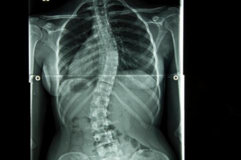 Chest X-ray and abdomen where one can appreciate the marked curvature of the spine (scoliosis)
Ultrasound After applying a cold always gel transmitter, the transducer or instrument which is placed on the area to be studied, emits ultrasound waves whose echo is transformed by the computer into a two or three dimensional image.
Although ultrasound is traditionally associated with monitoring of the fetus during pregnancy, it is also the main method of imaging the heart or one of the most important tests for the study of the brain of newborns and is essential for the diagnosis and study of the evolution of direct muscle injuries; that is, as a result of the shock of the muscle mass against a hard surface and the bone; They are common in contact sports and car accidents. And Indirect Muscle Injuries; they are injuries as a result of an intrinsic force generated by a sudden neurocontraction of the muscle (just to name one of the many etiologies). The most basic lesion is edema, which can translate into a focus of distention without fibrillar rupture (“contracture”) Or be present in any of the intrinsic forces injuries; stretches, strains, and different degrees of tears.
In the case of the lower limbs, ultrasound helps correct visualization of venous problems or blockages caused by blows or traumas and even tissue degeneration.
Patients very obese are more difficult to obtain ultrasound images because a larger amount of tissue attenuates (weakens) the sound waves as they penetrate deeper into the body and back to the transducer need for analysis.
Ultrasound has difficulty penetrating bone and therefore can only see the outer surface of bony structures and not what is inside (except infants because they have more cartilage in their skeletons older children or adults). To visualize the internal structure of bones or certain joints other imaging modalities such as RMA commonly used.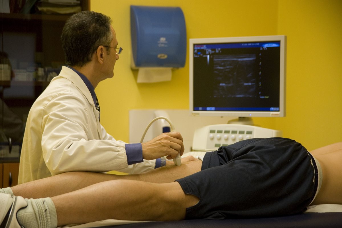
Doctor performing an ultrasound scan on hamstring
magnetic resonance (RMA)Normally when this test is performed prevents the patient has any metallic element, since the magnetic resonance generated images or dozens of sections of our anatomy using magnetic fields and radio waves.
These images are used "in many areas of medicine, such as neurological or abdominal diseases" with the intention of studying large volumes and different levels of a particular area of the body.
MRI is positioned as the test used in the detection and control of sports injuries. It is used for joint, ligament and tendon traumas so common in sports practices, and above all affecting the limbs.
Scanner or computed tomography (CT): : En esta prueba diagnóstica los rayos X se utilizan para generar imágenes transversales de nuestro cuerpo, reconstruidas por potentes ordenadores a partir de la información enviada por los receptores de radiación; es decir es un conjunto de emisión y captación, una interacción típica de ese tejido con la radiación electromagnética. “son rebanadas de nuestro cuerpo, como si nos cortasen como a una mortadela”, con la intención de observar múltiples patologías como cánceres o coágulos de sangre.

Computed tomography (CT)
Technology and Neurosciences
Magnetic resonance imaging (RMA), computed tomography (CT), electroencephalogram (EEG) and magnetoencephalography (MEG) and brain;
Thanks to these technologies, medicine has advanced by leaps and bounds in the investigation of neuroanatomy and in understanding the functioning of the brain. With the resonance it was possible to visualize the activity of the entire brain, thanks to the increase in blood flow, whose translation refers to the fact that different anatomical areas need nutrients to continue working: glucose and oxygen. On the other hand, thanks to the EEG "electroencephalogram" (the old method to detect brain waves) the MEG Magnetoencephalography was developed; ultra-precise recording of the tiny magnetic waves that accompany the discharge of currents in cortical neurons.
Con estas tecnologías se tejieron los correlatos neuronales de algunos fenomenos mentales que hasta finales de la decada de los ochenta eran confusos, y con muchos interrogantes . Se pudieron estudiar fenomenos como la recoleción de datos, las emociones, los sentimientos, los pensamientos y el sistema cognitivo (memoria,asociación, autorepresentación epacio/tiempo, toma de decisiones entre otras) . en En la actualidad estos fenómenos se encuentran muy bien explicados y ampliamente desarrollado por la ciencias que estudia la mente y el cerebro.
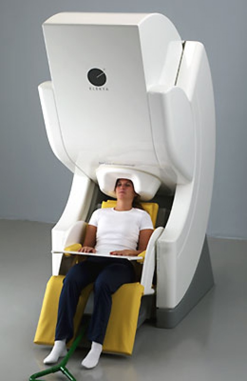
Magnetoencefalografía (MEG)
Conclusion:
As he stated in Article PROGRAM FOR Rehabilitative anterior cruciate ligament “the movement is the most beautiful risk of modern life”. Motion understand sports, dances, martial arts, circus activities, routine work, preparation and physical education.
Both movement and sedentary lifestyle and hypokinesia have risk factors; so that professionals who are in contact with these phenomena of contemporary life, undoubtedly have to know about the scope of these diagnostic technologies, which allow the detection and prevention of injuries, pain, trauma or pain; and consequently that these problems aggravate us. It is clear that for this purpose we must inquire about the daily life of the student/patient, but above all in the nature, sensation, location and duration of the pain, as well as the relationship that the student has had throughout of your life with this homeostatic feeling. However, as so many therapists and researchers of the body have taught us, there are osteoarticular pains whose cause is not on the point of pain, “…Evil is never found where the pain is manifested… “ Francoise Mezieres, estrategia de lectura corporal que se entrena y perfecciona con el correr de los años y los tratamientos terapéuticos y de entrenamientos empleados.A partir de esto queremos resaltar dos puntos principales:
1- That the scientific eye that each teacher/therapist acquires through study and experience is very important: That is, the observation of the body typology that would express certain sentimental and emotional states of our students.
Y 2- Que ese ojo científico puede ser agudizado y complementado con la exactitud fisicomatemática de las tecnologías diagnosticas y de los profesionales que las operan .
authors: Maria Jose Ghigo, BA in bioimaging. José De Laurentis, Bachelor of Physical Education.
SOURCES :
Websites: http://scielo.conicyt.cl
http: //entrenaenbarcelona.comtipos-de-lesiones-muscularis-and-treatment
Books:
“The diagnosis” Gian Franco Gramaccione -Paola Terrible.
“Muscle chains; lordosis, kyphosis and scoliosis”, Leopoldo Busquets.
“Anatomy for radiodiagnosis”, Ryan Mac Nicholas. Eustace.

")


